Advanced ultrasonography for accurate diagnosis and women’s health
High-precision imaging for comprehensive health assessment
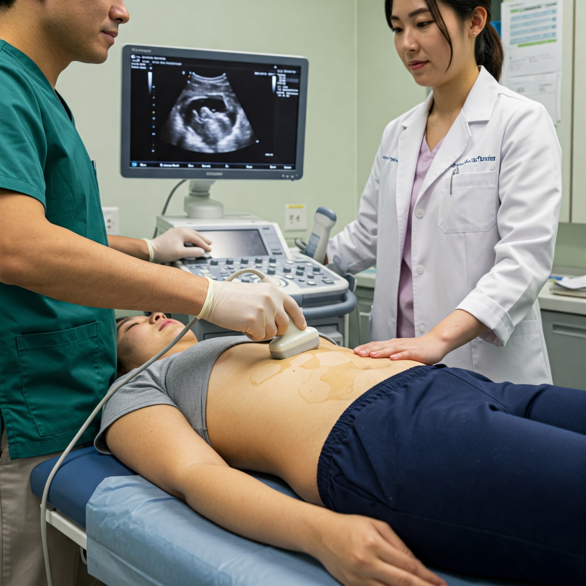
At Neo-Woman Hospital’s Ultrasonography Center, we provide state-of-the-art ultrasound examination services to support women’s health across all life stages. Our advanced imaging technology ensures accurate diagnosis, prenatal care, and early detection of health conditions with a patient-centric approach.
Our specialized ultrasonography in pregnancy offers expecting mothers detailed fetal screenings, anomaly scans, and growth monitoring, ensuring a healthy journey from conception to birth. We deliver comprehensive ultrasonography reports interpreted by expert radiologists for precise medical insights.
Looking for ultrasonography near me? At Neo-Woman Hospital, we provide safe, painless, and real-time imaging, helping detect gynecological conditions, assess reproductive health, and guide treatment plans effectively.
Many wonder about ultrasonography vs ultrasound—while the terms are often used interchangeably, ultrasonography refers to the technique, while ultrasound is the actual imaging. Regardless, our facility ensures both high-quality scans and expert interpretations for optimal healthcare outcomes.
With our cutting-edge technology and expert team, Neo-Woman Hospital’s Ultrasonography Center is committed to delivering precision, comfort, and reliable diagnostic support for every woman’s health journey.
Neo-diagnostics ultrasonography center
At Neo-Woman Hospital’s Neo-Diagnostics Center, we provide advanced ultrasonography services for accurate diagnosis and comprehensive women’s health care. Our expert radiologists and state-of-the-art imaging technology ensure precise and reliable ultrasound examinations, offering essential insights for early detection and treatment planning.
quality scans in a comfortable setting. Understanding ultrasonography vs ultrasound, ultrasonography refers to the technique of imaging, while ultrasound is the actual scan—at Neo-Diagnostics, we specialize in both, ensuring real-time, high-definition imaging for accurate health assessments.
From routine screenings to advanced diagnostic imaging, our Neo-Diagnostics Ultrasonography Center is committed to delivering precise results, expert care, and patient comfort, empowering women to make informed health decisions.
Whether you need ultrasonography in pregnancy to monitor fetal development or diagnostic imaging for gynecological conditions, our center delivers detailed ultrasonography reports with expert interpretations. If you’re searching for ultrasonography near me, our facility provides safe, high-
- The role of ultrasonography in women’s health
Ultrasonography plays a crucial role in diagnosing and monitoring various health conditions in women. As a non-invasive imaging technique, it allows doctors to assess reproductive organs, detect abnormalities, and monitor pregnancies without exposure to harmful radiation. From evaluating ovarian cysts and uterine fibroids to detecting endometriosis, ultrasonography is a valuable tool in gynecology.
It is also used to guide certain medical procedures, such as biopsies, ensuring precision and accuracy. For expecting mothers, fetal ultrasound provides critical insights into the baby’s development, helping detect congenital abnormalities and ensuring maternal well-being. With continuous advancements in imaging technology, ultrasonography remains one of the safest, most effective diagnostic methods for women’s health, enabling early detection and timely medical intervention.
Here are some of the check up and health screenings that you should get done:
- Pelvic Exam
- Pap Smear (Cervical Cancer Screening)
- Breast Exam
- Blood Pressure Measurement
- Cholesterol Screening
- Bone Density Scan (DXA)
- Blood Sugar Test (Glucose Screening)
- Thyroid Function Tests
- Blood Count (Complete Blood Count, CBC)
* The specific exams and screenings recommended can vary based on a woman’s age, family history, personal health history, and risk factors. It’s essential to discuss your individual healthcare needs and screening schedule with your healthcare provider.
- Ultrasonography in pregnancy: what every expecting mother should know
Ultrasonography in pregnancy is an essential tool that ensures the health and well-being of both the mother and the baby. Throughout pregnancy, ultrasound scans help monitor fetal growth, detect congenital abnormalities, and assess placental position. The first-trimester scan confirms the pregnancy and checks for any complications, while the mid-pregnancy anomaly scan provides a detailed view of the baby’s development. In the third trimester, ultrasounds help evaluate fetal position, amniotic fluid levels, and overall well-being. Doppler ultrasounds also assess blood flow to the fetus, ensuring proper oxygen and nutrient supply. Expecting mothers often search for “best ultrasonography in pregnancy services” to ensure they receive expert care. With modern advancements like 3D/4D ultrasound, parents can even see detailed images of their baby before birth.
- Ultrasonography vs other imaging techniques: which one is right for you?
Many people wonder about the difference between ultrasonography vs ultrasound and how it compares to other imaging techniques like MRI, CT scans, and X-rays. While ultrasonography refers to the medical technique of using sound waves to create images, ultrasound is the actual scan or image produced. Unlike X-rays and CT scans, which use radiation, ultrasonography is a safe, non-invasive method suitable for pregnant women and individuals needing frequent scans. Compared to MRI, ultrasound is faster, more cost-effective, and often performed in real time, allowing immediate diagnosis.
It is particularly useful in gynecology, cardiology, and abdominal imaging. Whether used for pregnancy monitoring or detecting organ abnormalities, ultrasonography remains a preferred diagnostic tool due to its safety, accessibility, and efficiency.
- Advancements in ultrasonography technology: the future of diagnostic imaging
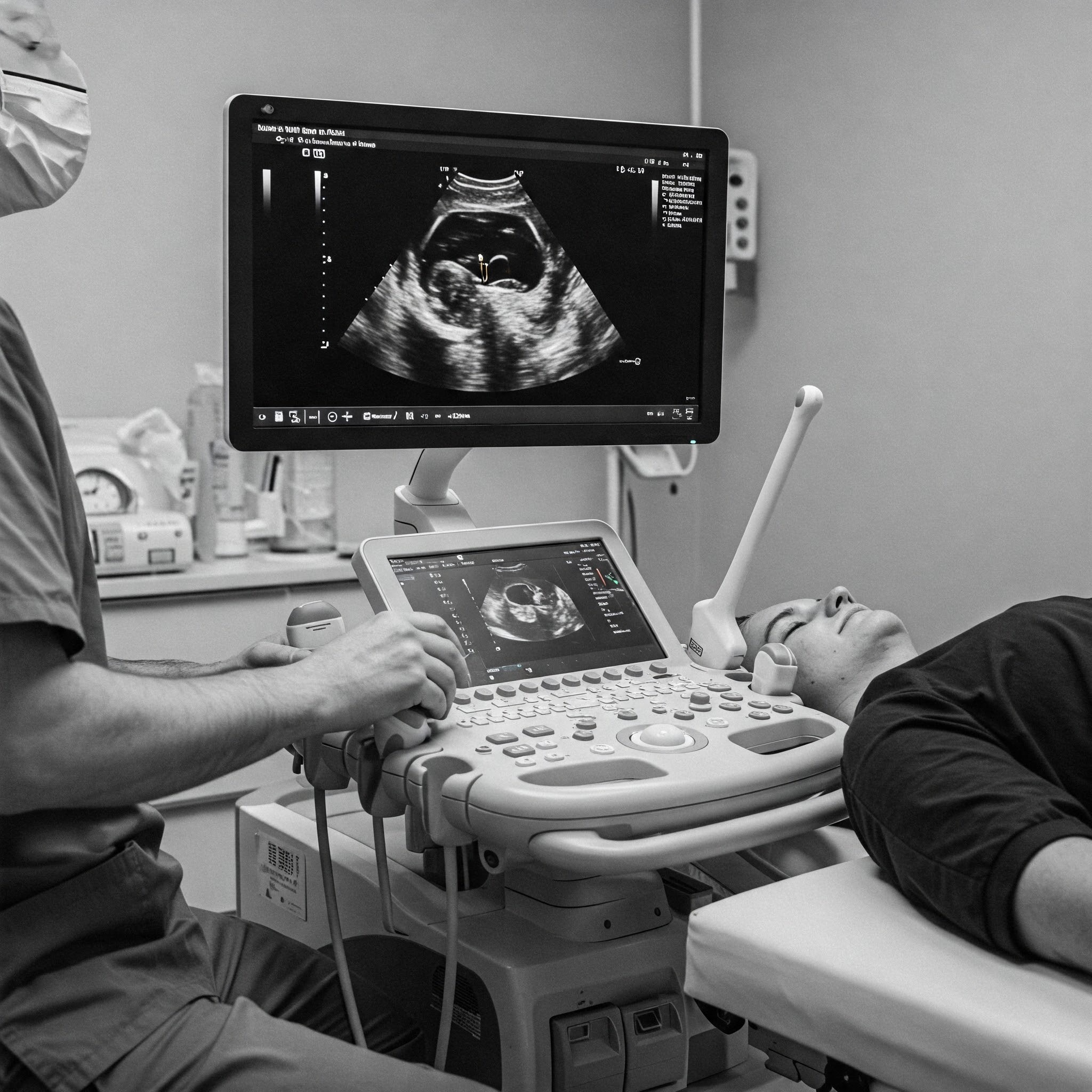
Ultrasonography has come a long way, evolving into a highly sophisticated ultrasound examination tool with advanced imaging capabilities. Traditional 2D ultrasound has now been complemented by 3D and 4D imaging, providing clearer and more detailed visuals of internal organs and fetal structures. Doppler ultrasound enables real-time assessment of blood flow, helping detect vascular conditions and fetal circulation issues.
AI-powered ultrasound technology is also emerging, allowing automated image analysis for faster and more accurate diagnoses. These innovations are making ultrasound examination even more reliable in detecting diseases early and guiding minimally invasive procedures. As the technology continues to improve, ultrasonography will remain a cornerstone of modern diagnostics, enhancing patient care and medical decision-making.
- Breaking myths about ultrasonography: facts every patient should know
There are many misconceptions surrounding ultrasonography, often causing unnecessary worry among patients.
One common myth is that ultrasound waves can be harmful, but research confirms that ultrasound is completely safe, as it uses sound waves instead of radiation. Another misconception is that all abnormalities will always be visible in an ultrasonography report, but in reality, some conditions require additional tests for confirmation.
Patients often assume that only pregnant women undergo ultrasound scans, whereas ultrasonography is widely used for diagnosing gynecological, cardiac, and abdominal conditions in both men and women. Some also believe that ultrasound results are immediate, yet complex cases require detailed analysis before final reporting. Understanding the facts behind ultrasonography helps patients make informed healthcare decisions with confidence.

Frequently asked questions
What is ultrasonography, and how does it work?
Ultrasonography is a non-invasive imaging technique that uses high-frequency sound waves to create real-time images of internal organs and tissues. A transducer emits these sound waves, which bounce off tissues and return as echoes, forming detailed visuals on a screen. This technology helps in diagnosing medical conditions, monitoring pregnancies, and guiding certain procedures.
Why is ultrasonography important during pregnancy?
Ultrasonography in pregnancy is crucial for monitoring fetal development, detecting abnormalities, and assessing the baby’s position, heartbeat, and growth. It ensures both maternal and fetal well-being by identifying any potential complications early, allowing timely medical intervention.
How do I prepare for an ultrasound examination?
Preparation varies depending on the type of ultrasound. For abdominal ultrasounds, fasting for 6–8 hours may be required, while ultrasound examination of the pelvis might require a full bladder for clearer imaging. Your doctor will provide specific instructions based on the type of scan you need.
What does an ultrasonography report include?
An ultrasonography report contains images from the scan along with a detailed analysis by a radiologist. It includes information about organ structures, abnormalities (if any), and relevant medical findings. The report helps doctors make accurate diagnoses and treatment plans.
Where can I find ultrasonography services near me?
If you’re searching for ultrasonography near me, Neo-Diagnostics offers advanced ultrasound imaging services with experienced radiologists and the latest technology for precise results.
Service line
Our locations
Neo-woman clinic
Apte Road
1212, Apte Rd, opp. Centro Hotel, Shirole Road, Shivajinagar, Pune, Maharashtra 411004
Contact us at: 9423039292
Our locations
Neo-woman clinic
Hadapsar
Gandharva Empire, 3rd floor, Above Fab India, Raskar Chowk, Amanora Park, Hadapsar, Pune, Maharashtra 411036
Contact us at: 7030114488
Copyright © 2026 neo-Woman – all rights reserved.
:::| powered by dimakh consultants |:::